You went through the surgery, endured the swelling, followed the soft-food diet, and assumed you were done with wisdom teeth forever. Then, months or even years later, you run your tongue along the back of your mouth and feel a hard, sharp bump pushing through the gumline.
A wave of dread sets in: Can wisdom teeth grow back?
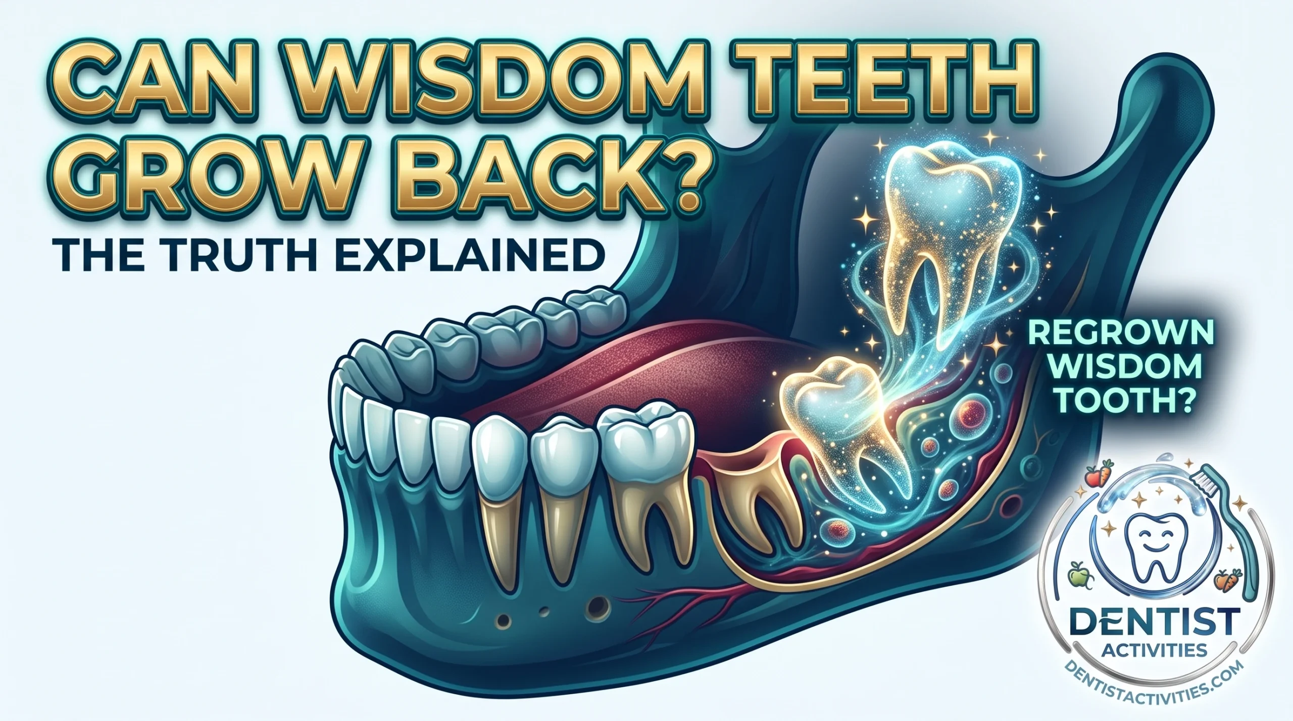
Having a third molar reappear feels like a cruel dental prank. The human body is full of surprises, but when it comes to teeth, biological rules are generally strict. In almost all cases, a tooth that has been fully extracted cannot regenerate. Yet thousands of dental patients experience symptoms that feel identical to a wisdom tooth emerging for a second time.
Here is a comprehensive look into human dental anatomy, the phenomenon of supernumerary teeth, common post-extraction quirks, and what is actually happening when you feel something hard erupting in the back of your jaw.
The Short Answer: Can Wisdom Teeth Really Grow Back?
Biologically speaking, a standard wisdom tooth cannot grow back once it has been surgically removed.
Humans are diphyodonts, meaning we develop two successive sets of teeth over our lifetimes: primary (baby) teeth and permanent (adult) teeth. Wisdom teeth—clinically known as third molars—are the last permanent teeth to develop and erupt, usually appearing between the ages of 17 and 25. Once an adult tooth is extracted, the body does not possess the stem-cell mechanism to spontaneously regrow a biological replacement in that empty socket.
However, many people genuinely do experience a “new” tooth or a tooth-like structure emerging in the exact site of an earlier extraction.
The Biological Reality of Permanent Teeth
Tooth development begins in the womb and continues through early adulthood via a specialized band of tissue called the dental lamina. This tissue creates a predetermined number of tooth buds:
- 20 primary teeth
- 32 permanent teeth (including 4 third molars)
Once a tooth bud develops, matures, and is later extracted, the dental lamina in that area is spent. The alveolar bone fills the socket, the gum tissue seals over it, and no new tooth tissue can form from scratch.
Why the Sensation of Regrowth Is So Common
If true biological regeneration is impossible, why do so many patients search “can wisdom teeth grow back” after feeling an unmistakable protrusion?
The answer typically falls into one of three categories:
- Hyperdontia: A genetic occurrence where your body created an extra (supernumerary) tooth bud behind the original wisdom tooth.
- Bone Sequestration: Small fragments of bone or hard tissue working their way out through the gums after surgery.
- Incomplete Extraction: A retained root tip or tooth fragment left behind during a complex extraction that has migrated toward the surface.
Supernumerary Teeth: The Science of Extra Third Molars
While teeth cannot regenerate, your body can produce more than the standard 32 adult teeth. This condition is known as hyperdontia, and the additional teeth are called supernumerary teeth.
What Are Hyperdontia and Distomolars?
When a supernumerary tooth develops directly behind a third molar, dental professionals refer to it as a distomolar (or a “fourth molar”). If an extra tooth forms adjacent to or alongside the molar, it is termed a paramolar.
Distomolars often look like miniature, rudimentary wisdom teeth, though some develop fully formed crowns and roots. If an oral surgeon removes your four standard wisdom teeth, an undiscovered distomolar hidden deeper within the jawbone may slowly migrate upward and erupt into the empty space years down the road. To the patient, it feels exactly like the original wisdom tooth grew back.
How Prevalent Are “Fifth” and “Sixth” Molars?
Hyperdontia is relatively uncommon, affecting roughly 1% to 4% of the global population, with a slightly higher prevalence in men than in women.
Supernumerary teeth can appear anywhere in the dental arch:
- Mesiodens: Extra teeth appearing between the two upper front teeth (the most common type).
- Distomolars: Extra molars appearing behind the third molars (the second most common type).
- Premolar region: Extra teeth forming between bicuspids.
Most distomolars remain impacted inside the jawbone and are discovered incidentally on panoramic dental X-rays. But when one has enough space and the correct eruptive path, it will push through the gum tissue just like a normal wisdom tooth.
Common Reasons It Feels Like Your Wisdom Teeth Are Growing Back
If you feel a hard, pointy, or irritating object in the back corner of your jaw after an extraction, an extra tooth is only one of several possibilities. More often, the culprit is a byproduct of normal bone remodeling or healing.
1. Bone Spicules and Bone Sequestra
The most frequent cause of post-extraction “tooth regrowth” is a bone spicule (also called a bone fragment or bone sequestrum).
During wisdom tooth extraction, the surrounding alveolar bone is subjected to pressure, sectioning, or manipulation. In the weeks and months following surgery, tiny slivers of dead or fractured bone may detach from the main jaw structure.
The body treats these detached bone slivers as foreign bodies. Through a natural expulsion process, the surrounding gum tissue slowly pushes the sharp fragment toward the surface. As it emerges:
- It feels hard and sharp against the tongue.
- It looks white or yellowish beneath the gums.
- It mimics the tip of an erupting crown.
In most cases, bone spicules work themselves completely free and fall out on their own, or your dentist can easily lift them away with minor topical numbing.
2. Incomplete Extraction (Retained Root Fragments)
Wisdom tooth roots can be long, curved, and intimately intertwined with sensitive structures like the inferior alveolar nerve in the lower jaw.
In certain complex cases, a surgeon may decide that fully extracting a deeply hooked or fragile root tip presents a high risk of permanent nerve damage. When this happens, the surgeon intentionally leaves a tiny, healthy piece of the root tip behind—a clinical approach related to a procedure called a coronectomy.
Over several years, the surrounding bone can push that small root remnant upward toward the gum surface. If it breaches the gumline, it creates the distinct impression of a re-emerging tooth.
3. Eruption or Shifting of Adjacent Second Molars
When wisdom teeth are extracted during the early teenage years, the adjacent second molars might not have finished settling into their final positions.
Without the crowded pressure of a third molar behind them, second molars may drift, tilt, or finish erupting at an angle that exposes hard cusp tips toward the back of the mouth. This structural realignment can feel like a new tooth breaking through.
4. Gingival Hyperplasia and Calcified Tissue
Occasionally, deep gum tissue undergoes localized scarring, cyst formation, or calcification following surgical trauma. A hardened pocket of fibrous scar tissue can mimic the firmness of an emerging tooth crown when pressed with a finger or tongue.
The Anatomy and Development of Wisdom Teeth
Understanding how wisdom teeth originate clarifies why they cannot simply regrow out of nowhere.
Why Do Humans Have Wisdom Teeth?
From an evolutionary perspective, our early human ancestors had larger, broader jaws and consumed a rough, abrasive diet consisting of raw meats, roots, fibrous plants, and nuts. This coarse diet caused significant dental wear and premature tooth loss. Third molars served as essential backup chewing power to ensure survival.
As human diets softened through agriculture and cooking, and our braincases expanded, our jaws evolved to become smaller. Today, modern human jaws frequently lack the spatial real estate required to accommodate 32 teeth, leading to impaction, misalignment, and the routine necessity of wisdom tooth extraction.
Dental Lamina and Tooth Bud Development
Tooth formation is an intricate process governed by genetic signaling:
- Initiation: Cells from the oral epithelium grow down into the underlying mesenchyme, forming the dental lamina.
- Bud Stage: Specialized groups of cells form distinct buds for each tooth.
- Cap and Bell Stages: The cells organize into the enamel organ, dental papilla, and dental follicle, laying down enamel and dentin.
- Crown and Root Formation: The visible crown mineralizes first, followed by the development of the roots, which push the tooth into the mouth.
Because this developmental cascade relies entirely on the initial presence of a genetic tooth bud, a new tooth cannot generate after the original structure is removed unless an extra bud was programmed into the jaw from the start.
Symptoms: Supernumerary Tooth vs. Normal Healing
Distinguishing between a genuine supernumerary tooth, a harmless bone spicule, and standard healing is key to deciding whether you need to visit an oral surgeon.
| Symptom / Feature | Supernumerary Tooth (Fourth Molar) | Bone Spicule (Sequestrum) | Post-Op Healing / Scar Tissue |
|---|---|---|---|
| Typical Onset | Months to years after surgery | 2 to 12 weeks post-surgery | 1 to 4 weeks post-surgery |
| Texture & Feel | Smooth, rounded, enamel-like cusp | Sharp, jagged, rough shard | Firm, slightly rubbery, or dull |
| Visual Appearance | White, tooth-shaped structure | Small, chalky white/yellow splinter | Red, pink, or pale scar tissue |
| Pain Level | Mild pressure or ache in the jaw | Minor prickling or tongue irritation | Tender to pressure; improves weekly |
| Mobility | Rigidly anchored into the jawbone | May become loose as it exits gums | Attached to soft tissue |
| Resolution | Requires monitoring or extraction | Sloughs off naturally or is removed | Softens and remodels over time |
How Dentists Diagnose Phantom Wisdom Teeth
If you suspect something is growing in your extraction site, a professional evaluation involves specific diagnostic steps to determine the exact cause.
1. Clinical and Tactile Examination
Your dentist will visually inspect the area for inflammation, gum redness, or visible white margins. Using a dental probe, they gently check the density and mobility of the protrusion. A loose, sharp fragment indicates a bone spicule, while a solid, anchored surface suggests a tooth crown or retained root.
2. Panoramic Radiographs (OPG)
A standard panoramic X-ray captures your entire upper and lower jaw on a single flat image. This scan instantly confirms:
- Whether an unsuspected fourth molar is present inside the bone.
- Whether a retained root fragment is migrating.
- The condition of the surrounding jawbone density.
3. Cone Beam Computed Tomography (CBCT)
If an extra tooth or complex root fragment is detected near a major anatomical pathway—such as the maxillary sinus or the mandibular nerve canal—a 3D CBCT scan provides volumetric imaging. This reveals the precise millimeter-level relationship between the extra tooth and critical nerves, ensuring any necessary removal is safe and predictable.
Treatment Options If an Extra Wisdom Tooth Emerges
If diagnostic imaging confirms that you have an erupting fourth molar, your treatment plan will depend on its position, alignment, and impact on your oral health.
When Extraction Is Necessary
Just like standard third molars, supernumerary molars often run out of room. Extraction is recommended if the extra tooth causes:
- Pericoronitis: Chronic bacterial infection of the gum flap covering a partially erupted crown.
- Damage to Adjacent Teeth: Pressure resorption on the roots of the neighboring second molar.
- Cyst Formation: Fluid-filled follicular sacs around an impacted crown that can hollow out jawbone tissue.
- Orthodontic Relapse: Crowding that pushes straight teeth out of alignment.
- Sinus Complications: Upper distomolars impinging upon the floor of the maxillary sinus.
When Observation (Monitoring) Is the Best Approach
If a fourth molar is small, completely embedded deep inside dense bone, asymptomatic, and not threatening any nerve pathways or adjacent roots, the most conservative approach is periodic monitoring. Your dentist will evaluate the tooth with routine X-rays every few years to confirm it remains dormant.
What to Do If You Feel Something Hard in Your Gums Post-Extraction
Finding an unexpected hard protrusion in your mouth can be unsettling, but taking the correct steps prevents infection and complications:
- Avoid Poking with Tools: Do not attempt to dig, pry, or scrape the area with toothpicks, tweezers, or fingernails. This introduces oral bacteria into healing tissue and risks infection.
- Use Warm Salt Water Rinses: Dissolve half a teaspoon of salt into a glass of warm water. Gently rinse the area after meals to keep the tissue clean and soothe localized inflammation.
- Monitor with Your Tongue (Gently): Take note of whether the object is sharp and mobile or fixed and broad.
- Schedule a Dental Checkup: Contact your dentist or oral surgeon for a quick exam and X-ray. If it is a minor bone spicule, they can remove it in seconds with virtually no discomfort.
Frequently Asked Questions
Can wisdom teeth grow back after being fully pulled?
No, a wisdom tooth that has been completely extracted cannot regenerate. Humans only develop one set of permanent molars. If a new tooth emerges in that spot, it is an extra, supernumerary tooth (fourth molar) that developed from a separate tooth bud.
How common is it to have extra wisdom teeth?
Supernumerary molars (distomolars) occur in approximately 1% to 2% of the general population. Many people never realize they have them unless they show up on a panoramic X-ray or begin to erupt later in life.
Why do I feel a sharp, white shard in my gums after wisdom tooth surgery?
This is almost always a bone spicule (bone sequestrum)—a microscopic fragment of jawbone that detached during the extraction. Your gums naturally push this fragment out as the tissue heals. It is harmless and can be removed easily by your dentist if it does not fall out on its own.
Can wisdom teeth grow back in your 30s, 40s, or 50s?
Standard wisdom teeth cannot grow back at any age. However, a dormant, impacted supernumerary tooth or a previously unnoticed root fragment can slowly shift and erupt through the gums decades after your initial surgery due to natural bone remodeling and age-related gum recession.
Is removing a fourth molar harder than a regular wisdom tooth?
The difficulty depends on its size, root anatomy, and location. Many distomolars are smaller and have simpler, fused roots compared to full-sized third molars, often making extraction straightforward. However, if the tooth is deeply impacted or close to a nerve, specialized surgical care is required.
While discovering a hard bump at an old extraction site can feel alarming, true wisdom teeth cannot regenerate once removed. In the vast majority of cases, what feels like a re-emerging tooth is simply a minor bone spicule working its way free, or, in rare cases, a harmless extra tooth bud that nature added to your jaw. A brief visit to your dentist and a routine X-ray will quickly demystify the phantom tooth and provide the right solution to keep your smile healthy and comfortable.